Home
/ Posterior View Of Heart Coronary Arteries - Illustrated Guide To Anomalous Coronary Activity An Interactive 3d Model Ctisus Com Ct Scanning - These 3d models of perfusion fixed human hearts display the varying anatomy of the coronary artery system.
Posterior View Of Heart Coronary Arteries - Illustrated Guide To Anomalous Coronary Activity An Interactive 3d Model Ctisus Com Ct Scanning - These 3d models of perfusion fixed human hearts display the varying anatomy of the coronary artery system.
Posterior View Of Heart Coronary Arteries - Illustrated Guide To Anomalous Coronary Activity An Interactive 3d Model Ctisus Com Ct Scanning - These 3d models of perfusion fixed human hearts display the varying anatomy of the coronary artery system.. The left main coronary artery gives rise to the left anterior descending artery and the left circumflex coronary artery. The 2008 american heart association/american college of cardiology guidelines give a class i recommendation for surgical coronary revascularization in patients with acaos and any of the following : This process is called atherosclerosis. Front view (anterior) of the heart outside view of the back (posterior) of the heart. Complete visualization of these arteries and their branches requires care and rigor to ensure complete anatomical documentation.
Together with the left anterior descending artery, the right coronary artery helps supply blood to the middle or septum of the heart. Each artery is a muscular tube lined by smooth tissue and has three layers: Coronary artery disease (cad) coronary artery disease is caused by plaque buildup in the wall of the arteries that supply blood to the heart (called coronary arteries). Anomalous left main coronary artery coursing between the great arteries; Plaque is made up of cholesterol deposits.
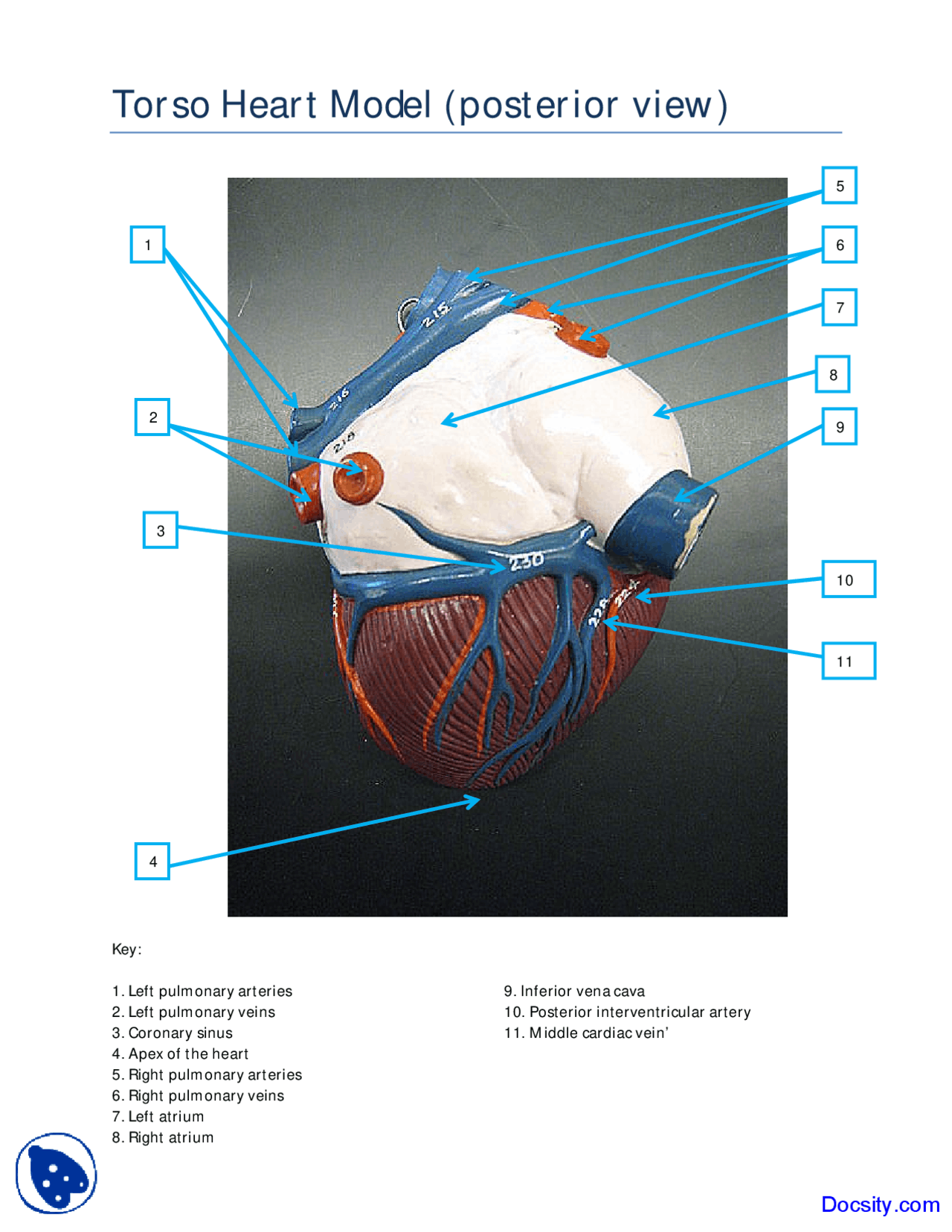
Posterior View Of Torso Heart Model Human Anatomy Handout Docsity from static.docsity.com A complete blockage can cause a heart attack. In general, coronary arteries traverse the surface of the heart and are encased in varying amounts of epicardial fat. The rest of the right coronary artery and its main branch, the posterior descending artery, together with the branches of the circumflex artery, run across the surface of the heart's underside, supplying the bottom portion of the left ventricle and back of the septum. The left main coronary artery gives rise to the left anterior descending artery and the left circumflex coronary artery. The anterior cardiac veins parallel the small cardiac arteries and drain the anterior surface of the right ventricle. The rca normally arises from the right coronary sinus (cs) and courses in the right av groove toward the crux of the heart (the point on the posterior surface of the heart where the av groove transects the line of the interventricular septum and interatrial septum, forming a cross). A contrast dye was injected through the right and left coronary ostia into the coronary arteries. The coronary arteries supply blood, oxygen and nutrients to your heart.
Each artery is a muscular tube lined by smooth tissue and has three layers:
We will discuss cardiac physiology, incorporating blood flow through the heart, coronary systole and diastole, the events that occur during The coronary arteries also include the collateral coronary arteries, small blood vessels that connect the normal coronary arteries with one another. The right coronary artery divides into smaller branches, including the right posterior descending artery and the acute marginal artery. Treats blocked heart arteries by taking arteries or veins from other parts of your body — called grafts — and using them to reroute the blood around the clogged artery to supply blood flow to your heart muscle. View an illustration of coronary bypass (link opens in new window). Front view (anterior) of the heart outside view of the back (posterior) of the heart. A contrast dye was injected through the right and left coronary ostia into the coronary arteries. On the posterior surface of the heart, the right coronary artery gives rise to the posterior interventricular artery, also known as the posterior descending artery. The rest of the right coronary artery and its main branch, the posterior descending artery, together with the branches of the circumflex artery, run across the surface of the heart's underside, supplying the bottom portion of the left ventricle and back of the septum. A posterior wall mi occurs when posterior myocardial tissue (now termed inferobasilar), usually supplied by the posterior descending artery — a branch of the right coronary artery in 80% of. The lad is one of two major branches of the lmca, with the other being the left circumflex (lcx) coronary arteries. These 3d models of perfusion fixed human hearts display the varying anatomy of the coronary artery system. Documented coronary ischemia secondary to coronary artery compression when.
View an illustration of coronary bypass (link opens in new window). The anterior cardiac veins parallel the small cardiac arteries and drain the anterior surface of the right ventricle. A contrast dye was injected through the right and left coronary ostia into the coronary arteries. These 3d models of perfusion fixed human hearts display the varying anatomy of the coronary artery system. The left coronary artery (lca) arises from the left posterior aortic sinus and quickly bifurcates into the left circumflex artery (lcx) and left anterior descending artery (lad), which supply blood to the left atrium and left ventricle.
Clinical Anatomy Cardiac Coronary Vessels Left And Right Coronary Artery Venous Sinus Youtube from i.ytimg.com The picture (above) shows the rca and the lad arteries. The aim of this paper was to describe color doppler imaging and flow reserve of the pd, regardless of its origin from the right or circumflex coronary artery, in different settings such as acute. Each artery is a muscular tube lined by smooth tissue and has three layers: The high mortality associated with cad makes the development of medical interventions that repair and replace diseased arteries a high priority for. When the heart is healthy, these vessels play only a minor role. Eventually, the reduced blood flow may cause chest pain (angina), shortness of breath, or other coronary artery disease signs and symptoms. Dominance of one side over the other is determined by which artery system supplies the posterior descending artery, which supplies the backside of the heart as well as the interventricular septum (the thick wall that separates the heart's ventricles, or lower chambers). The coronary arteries also include the collateral coronary arteries, small blood vessels that connect the normal coronary arteries with one another.
Plaque buildup causes the inside of the arteries to narrow over time.
View an animation of blood flow (link. Complete visualization of these arteries and their branches requires care and rigor to ensure complete anatomical documentation. The rest of the right coronary artery and its main branch, the posterior descending artery, together with the branches of the circumflex artery, run across the surface of the heart's underside, supplying the bottom portion of the left ventricle and back of the septum. When the heart is healthy, these vessels play only a minor role. The lad, the cx, and the rc. The 2008 american heart association/american college of cardiology guidelines give a class i recommendation for surgical coronary revascularization in patients with acaos and any of the following : This process is called atherosclerosis. The lm artery quickly branches into two large arteries — the left anterior descending artery (lad) and the circumflex artery (cx). A posterior wall mi occurs when posterior myocardial tissue (now termed inferobasilar), usually supplied by the posterior descending artery — a branch of the right coronary artery in 80% of. The picture (above) shows the rca and the lad arteries. The lad is one of two major branches of the lmca, with the other being the left circumflex (lcx) coronary arteries. Dominance of one side over the other is determined by which artery system supplies the posterior descending artery, which supplies the backside of the heart as well as the interventricular septum (the thick wall that separates the heart's ventricles, or lower chambers). These 3d models of perfusion fixed human hearts display the varying anatomy of the coronary artery system.
A contrast dye was injected through the right and left coronary ostia into the coronary arteries. The anterior cardiac veins parallel the small cardiac arteries and drain the anterior surface of the right ventricle. When the heart is healthy, these vessels play only a minor role. The rca normally arises from the right coronary sinus (cs) and courses in the right av groove toward the crux of the heart (the point on the posterior surface of the heart where the av groove transects the line of the interventricular septum and interatrial septum, forming a cross). Complete visualization of these arteries and their branches requires care and rigor to ensure complete anatomical documentation.
Heart Amboss from media-us.amboss.com This process is called atherosclerosis. The coronary arteries also include the collateral coronary arteries, small blood vessels that connect the normal coronary arteries with one another. The aim of this paper was to describe color doppler imaging and flow reserve of the pd, regardless of its origin from the right or circumflex coronary artery, in different settings such as acute. The heart muscle itself, then, is supplied by one of these three major coronary arteries: The lad, the cx, and the rc. Coronary artery disease (cad) coronary artery disease is caused by plaque buildup in the wall of the arteries that supply blood to the heart (called coronary arteries). Together with the left anterior descending artery, the right coronary artery helps supply blood to the middle or septum of the heart. The rca normally arises from the right coronary sinus (cs) and courses in the right av groove toward the crux of the heart (the point on the posterior surface of the heart where the av groove transects the line of the interventricular septum and interatrial septum, forming a cross).
Dominance of one side over the other is determined by which artery system supplies the posterior descending artery, which supplies the backside of the heart as well as the interventricular septum (the thick wall that separates the heart's ventricles, or lower chambers).
Treats blocked heart arteries by taking arteries or veins from other parts of your body — called grafts — and using them to reroute the blood around the clogged artery to supply blood flow to your heart muscle. Eventually, the reduced blood flow may cause chest pain (angina), shortness of breath, or other coronary artery disease signs and symptoms. Complete visualization of these arteries and their branches requires care and rigor to ensure complete anatomical documentation. View an illustration of coronary bypass (link opens in new window). In general, coronary arteries traverse the surface of the heart and are encased in varying amounts of epicardial fat. The anterior interventricular sulcus is visible on the anterior surface of the heart, whereas the posterior interventricular sulcus is visible on the posterior surface of the heart. Front view (anterior) of the heart outside view of the back (posterior) of the heart. The rest of the right coronary artery and its main branch, the posterior descending artery, together with the branches of the circumflex artery, run across the surface of the heart's underside, supplying the bottom portion of the left ventricle and back of the septum. The rca normally arises from the right coronary sinus (cs) and courses in the right av groove toward the crux of the heart (the point on the posterior surface of the heart where the av groove transects the line of the interventricular septum and interatrial septum, forming a cross). The picture (above) shows the rca and the lad arteries. The aim of this paper was to describe color doppler imaging and flow reserve of the pd, regardless of its origin from the right or circumflex coronary artery, in different settings such as acute. The right coronary artery divides into smaller branches, including the right posterior descending artery and the acute marginal artery. Located between the left and right ventricles are two additional sulci that are not as deep as the coronary sulcus.

